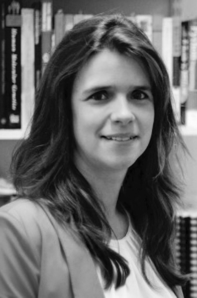
With a background in Computer Science, Claudia Lindner has been with the Centre for Imaging Sciences since 2010. After gaining her PhD, Claudia became a Research Associate in 2014 and has been working in the field of Medical Image Computing.
Posted on April 5, 2018

“I was recently awarded an Ernest Rutherford Fellowship, which is supported by UK Research and Innovation (UKRI) and funded by Health Data Research UK (HDR UK).
I will be doing research on developing and clinically validating an automated computer-aided system for analysing the efficacy of knee replacement surgery. The system to be developed will be based on the BoneFinder®-technology to outline the knee (bones and implants) in radiographs. The automatically identified structures will then be used to quantify the implant fitting to identify radiographic indications of early joint failure, informing the KRS decision-making process in knee replacement surgery and the resulting medical management.
There are an increasing number of medical images being gathered in all UK NHS hospitals, with the growing need and opportunity to utilise this information to improve the health of the nation. Currently, medical images are underutilised in clinical practise and research into musculoskeletal diseases where 2D radiographs are the imaging technique of choice due to wide availability, speed of acquisition and low cost. This three year fellowship will give me the opportunity to learn about how data is accessed and used within the Connected Health Cities (CHC) network, and how health interventions are rolled out in the NHS through CHC and HeRC programmes. During the fellowship, I will acquire the skills and knowledge needed to integrate medical image computing technologies with broader health informatics approaches.
I believe there are many opportunities in clinical practice of where automated image analysis techniques will be beneficial in assisting the clinical decision-making process. So it is fantastic to be able to gain a deeper understanding on how this can be achieved and to establish collaborations that will be beneficial for bringing advances in medical image computing into clinical practice.
"I have been developing robust and accurate systems for locating the outlines of bones and other structures in widely used medical images such as radiographs. My long-term goal is to develop digital health care solutions that lead to improvements in clinical practice."
A big focus of this project is to learn about how to get the technology, a computer-aided system, into clinical practice using the existing digital healthcare infrastructure. To complement my technical background, I will undertake several placements in a clinical environment to better understand clinical workflows, identify clinical needs for software solutions, explore new research avenues, and investigate the feasibility of introducing such a computer-aided system into clinical practice.
During my PhD research, I have developed a software system, BoneFinder® (www.bone-finder.com), to robustly and accurately outline the bone contours of the hip in 2D radiographs. BoneFinder® has been shown to be the best yet published automated system for this task. The performance of BoneFinder® is world-leading for numerous structures, and was awarded the first prize in an international ISBI Grand Challenge on Dental Image Analysis in 2015. BoneFinder® is being used by researchers world-wide, and the underlying BoneFinder®-technology has been patented and commercialised in a range of products.
My fellowship project will utilise the underlying BoneFinder®-technology to develop a computer-aided system for analysing the efficacy of knee replacement surgery by identifying radiographic signs of early joint failure. A feasibility study will be conducted to identify the clinical requirements for usability and acceptability, and for the integration of the system into the clinical workflow. The aim is for the system to be applied to the routinely collected radiographs of subjects with pain and disability in the knee who undergo knee replacement surgery. The system will assist the decision-making process in knee replacement surgery to identify loose or damaged implants before irreversible harm is done to the knee joint. The overarching goal is to transform clinically collected image data into useful medical information to benefit healthcare at individual and societal levels.